|
 Wednesday, December 1, 2010 Wednesday, December 1, 2010 |
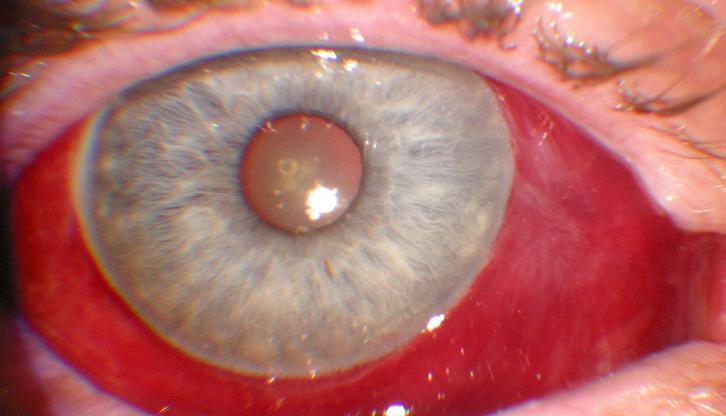
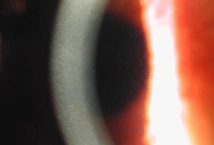
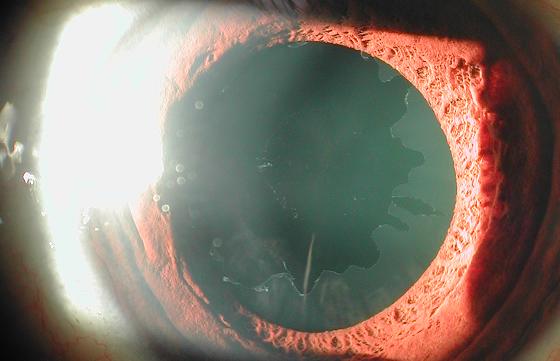
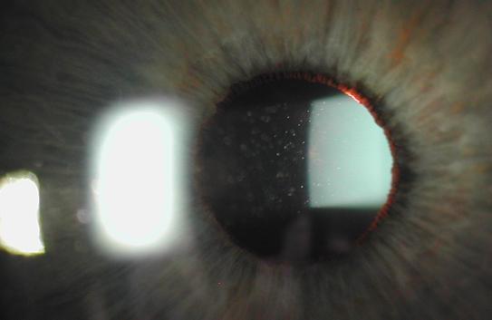
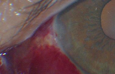
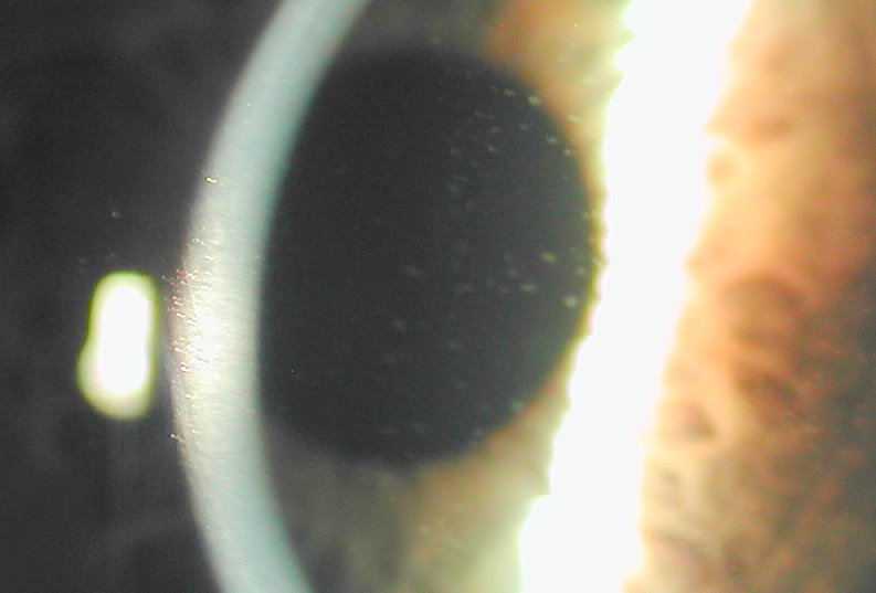
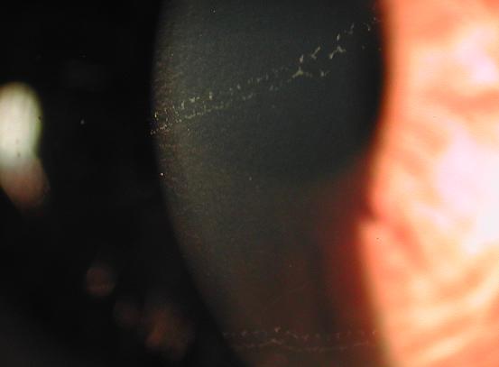
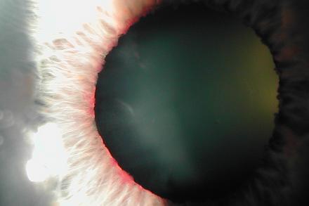
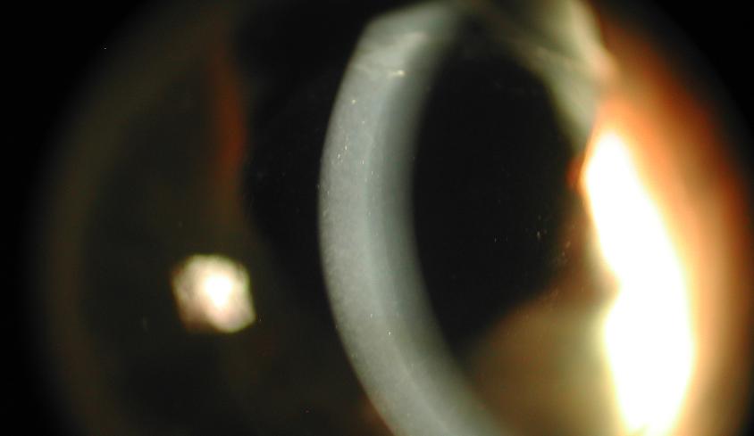
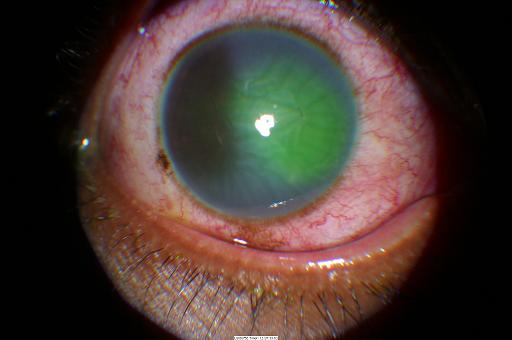
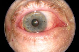
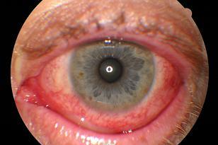
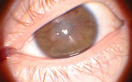
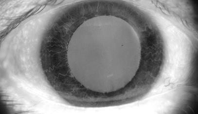
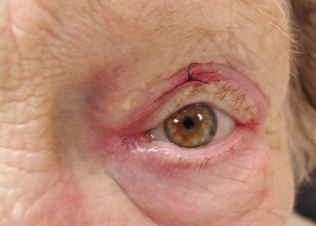
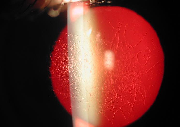
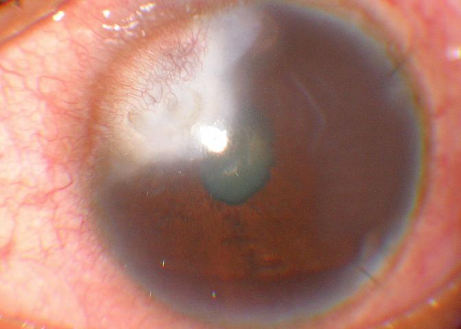
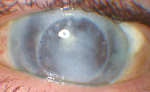
| Epithelial rejection line |
|
|
|
|
|
Tuesday, October 12, 2010 |
|
|
|
Tuesday, October 12, 2010 |
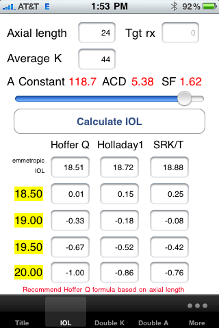
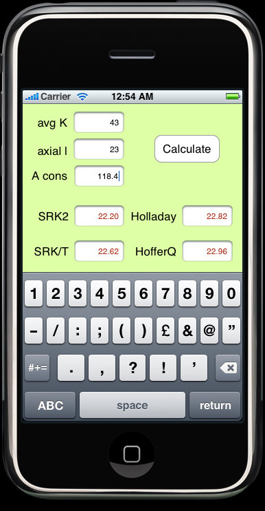
| The new Hoffer Programs version 3 for iPhone |
|
disclosure: I have a financial interest in this software.

|
alsobol at 1:28 PM | | |
Permalink
|
|
|
|
Tuesday, October 12, 2010 |
|
|
|
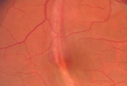
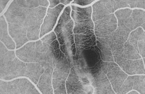
Friday, May 7, 2010 |
| The picture tells a story. |
|
|
|
|
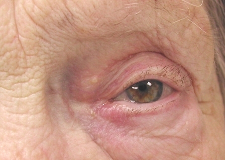
Friday, May 7, 2010 |
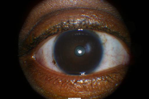
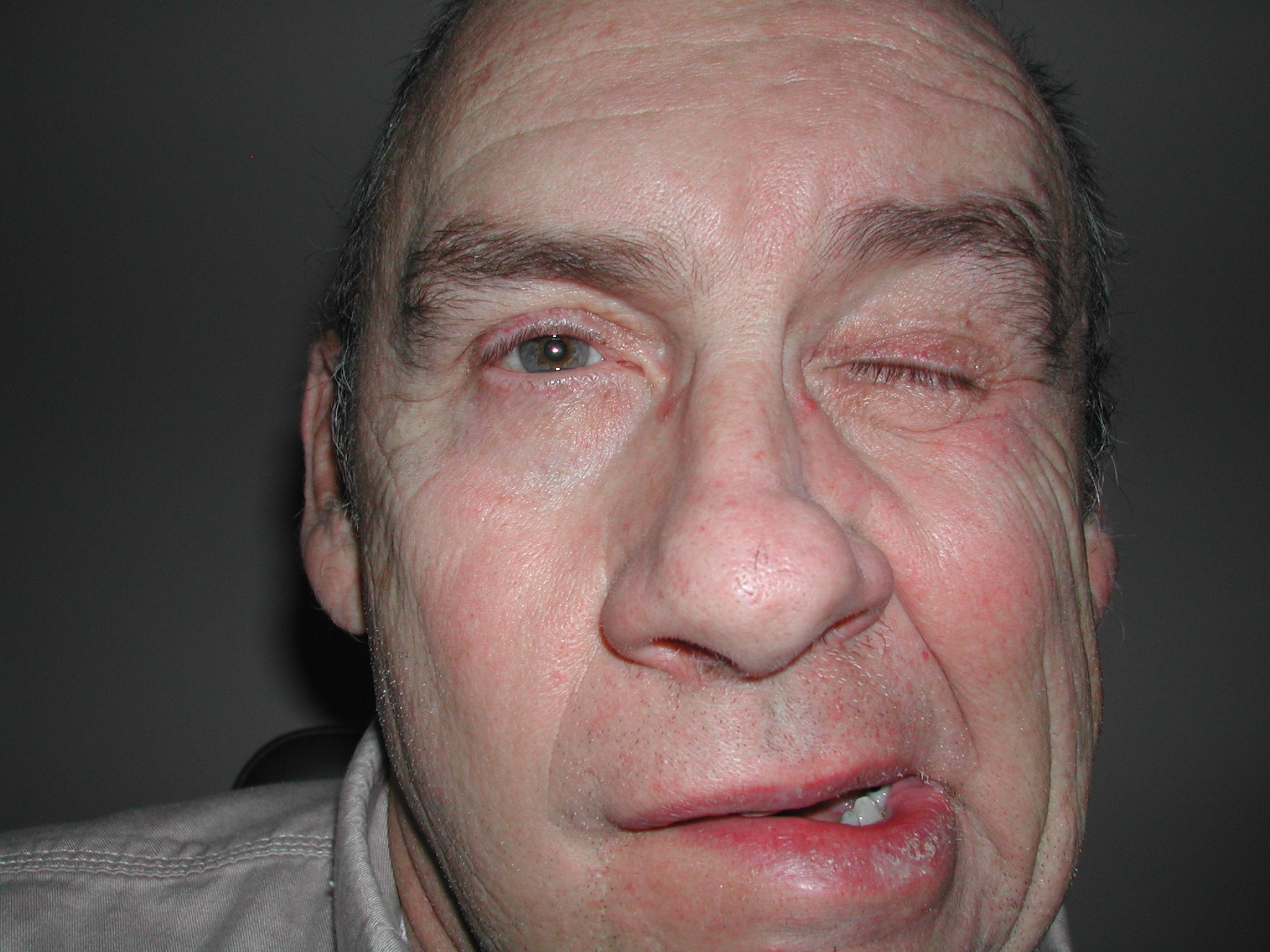
I haven't seen this since medical school and saw two cases in two months. One was definitely with the chromosome deletion and the other was based on the phenotype.
|
alsobol at 9:06 AM | | |
Permalink
|
|
|
|
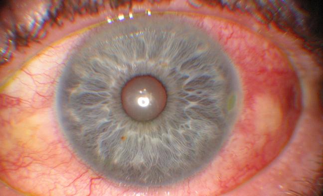
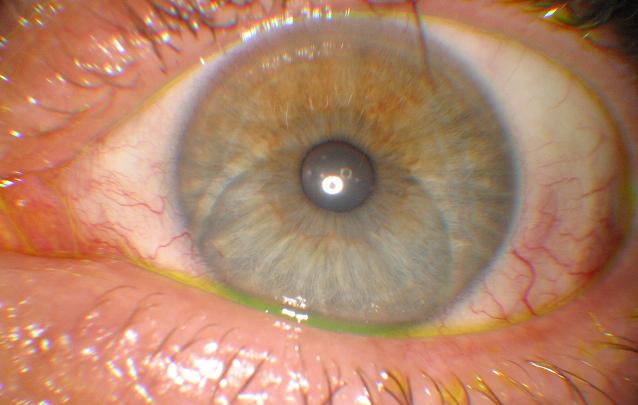
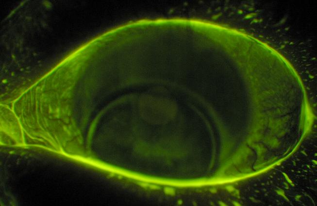
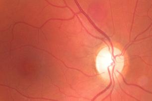
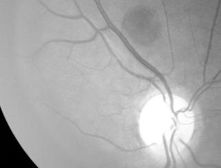
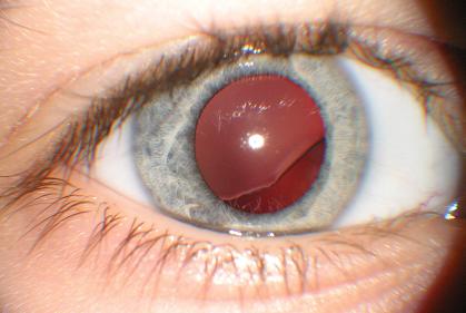
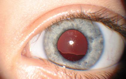
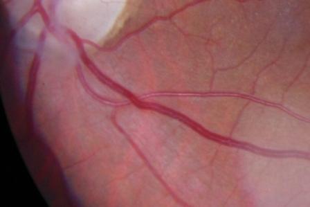
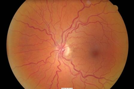
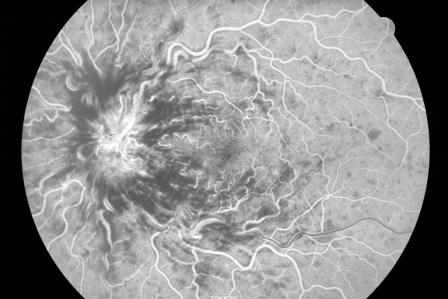
Friday, May 7, 2010 |
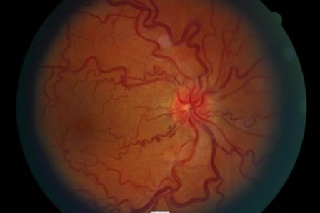
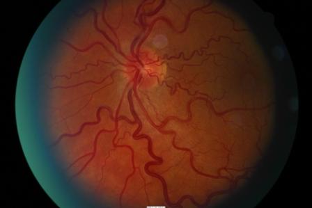
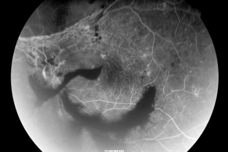
Haven't posted in awhile but cases keep coming in. I saw two young patients with CRVO's in the past two weeks. One has associated PIRW. The other was a young patient with severe stenosis.
Here they are:
|
alsobol at 9:03 AM | | |
Permalink
|
|
|
|
Wednesday, March 3, 2010 |
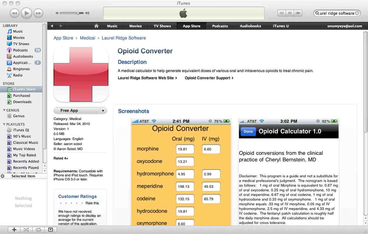
| First program on the app store |
|
My first app was approved today. It's an opioid converter for the iPod or iPhone. Pretty cool, eh? Not searchable yet but it's there.
|
alsobol at 5:00 PM | | |
Permalink
|
|
|
|
Wednesday, February 24, 2010 |
|
|
|
Friday, January 22, 2010 |
|
|
|
Friday, January 22, 2010 |
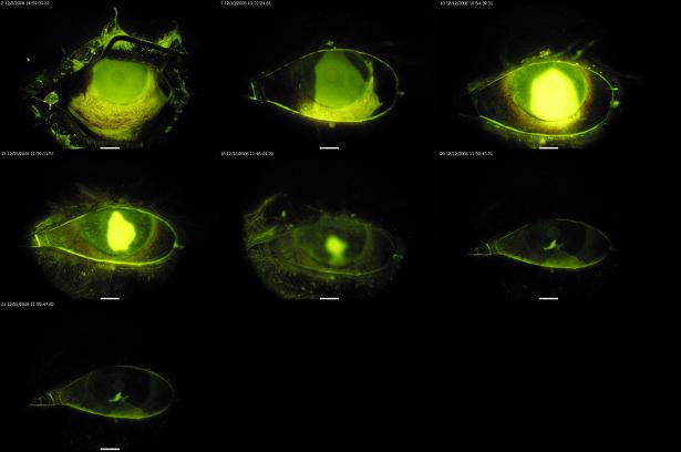
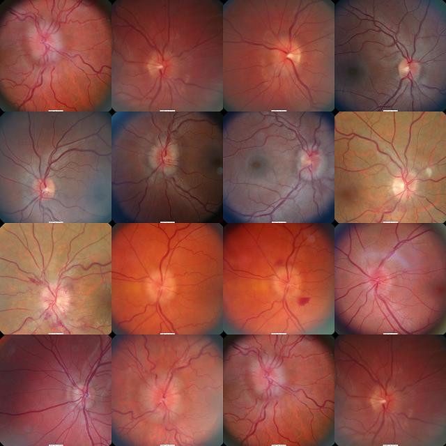
| Myelinated nerve fiber level collage |
|
|
|
|
Friday, January 22, 2010 |
|
|
|
Saturday, December 12, 2009 |
|
|
|
Saturday, December 12, 2009 |
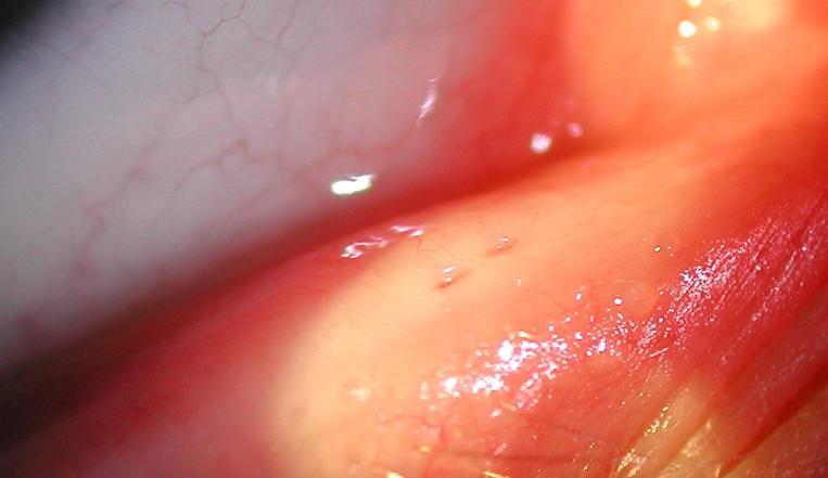
| Nodular episcleritis with dellen formation |
|
This is my fourth blog post with a picture of a corneal dellen. I don't know what it is about these things that makes them so interesting to me.
|
alsobol at 6:42 AM | | |
Permalink
|
|
|
|
Friday, November 20, 2009 |
| Corneal indentation from RGP |
|
Luckily the groove isn't in the visual axis.
|
alsobol at 10:13 AM | | |
Permalink
|
|
|
|
Wednesday, October 21, 2009 |
| testing color vision filters |
|
|
|
|
Friday, October 16, 2009 |
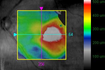
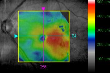
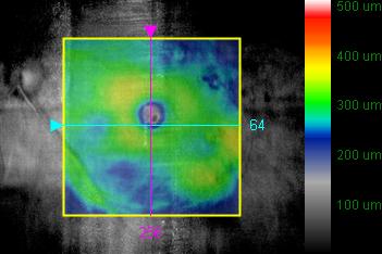
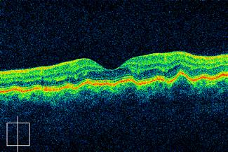
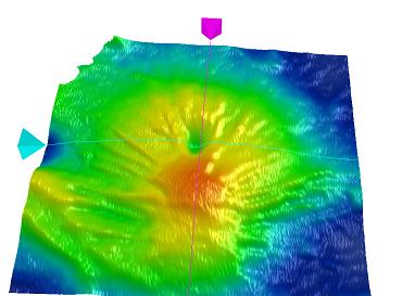
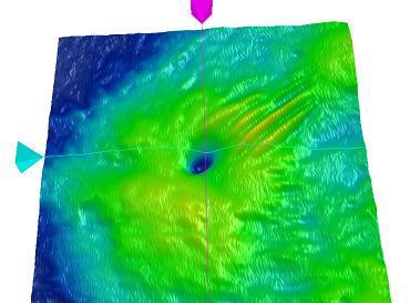
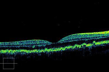
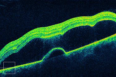
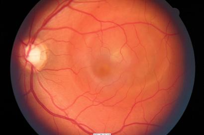
| Picture perfect resolution of macular edema |
|
Resolution of diabetic macular edema after a single focal laser treatment. The second OCT is at two months postop and the final one is at eight months.
|
alsobol at 12:30 PM | | |
Permalink
|
|
|
|
Tuesday, October 6, 2009 |
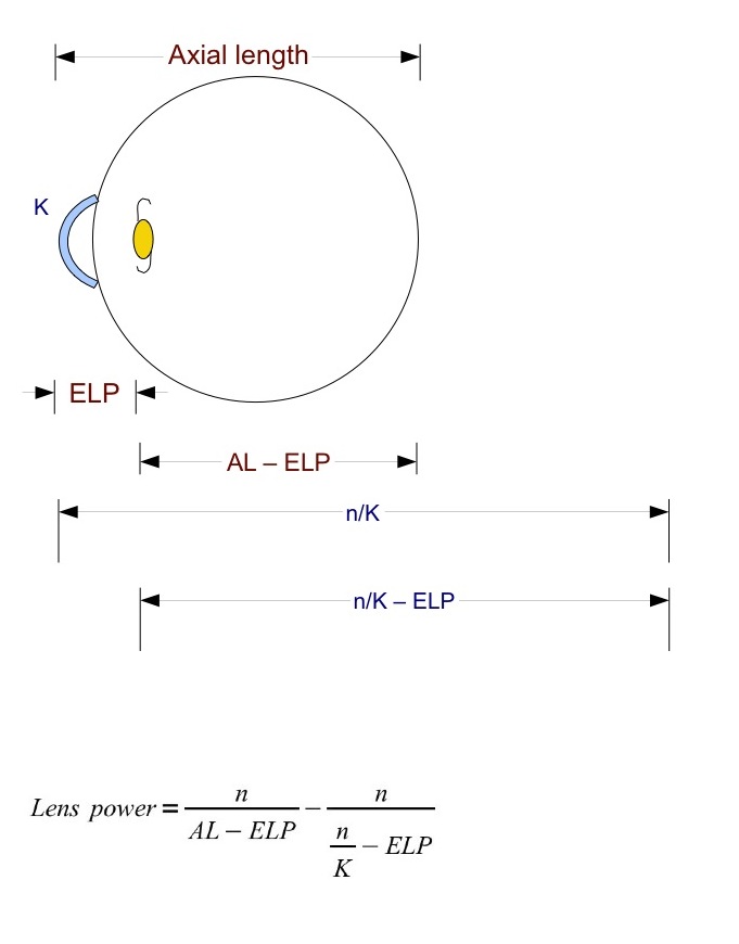
| Fyodorov equation derivation |
|
Worked on this all day; forgot how to add lens vergences. It's for an ELP program I'm writing.
|
alsobol at 7:32 PM | | |
Permalink
|
|
|
|
Tuesday, August 25, 2009 |
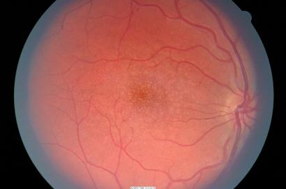
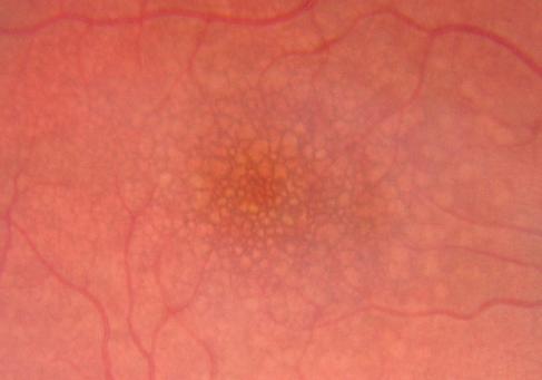
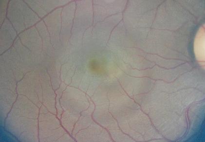
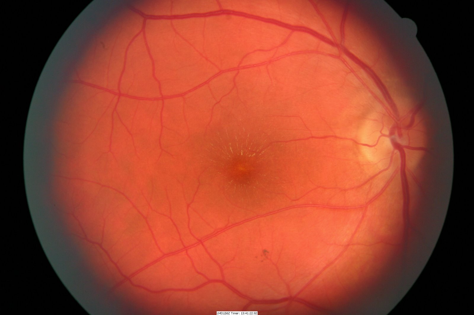
| More idiopathic choroidal striae |
|
I saw two cases of this last year, too. They are all bilateral and kinda interesting.
|
alsobol at 1:05 PM | | |
Permalink
|
|
|
|
Tuesday, August 25, 2009 |
|
|
|
Tuesday, July 14, 2009 |
| More Xcode stuff for the iPhone |
|
I'm still working on and off on an iPhone app. Here's how far I've gotten so far.
|
alsobol at 7:33 PM | | |
Permalink
|
|
|
|
Friday, July 10, 2009 |
| Local macular hole epidemic |
|
Lot of these macular holes over the past two weeks. Kind of unusual for a solo guy in general practice. I hit for the cycle today, having seen a pseudohole and stages 1 through 4. Here are a few of them: Pseudohole 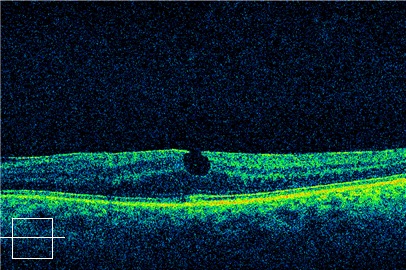 Stage 1 hole 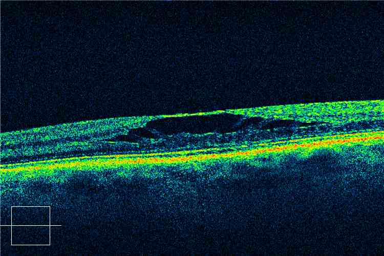 Stage 2 Hole 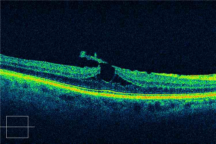 Stage 3 Hole 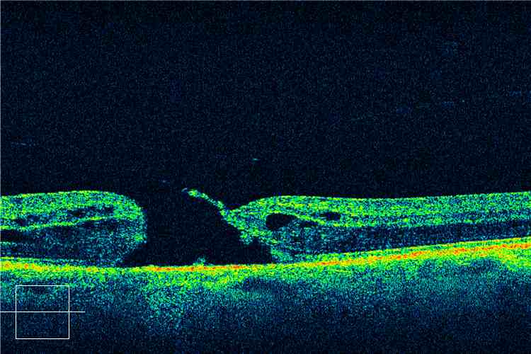
|
alsobol at 1:17 PM | | |
Permalink
|
|
|
|
Tuesday, June 16, 2009 |
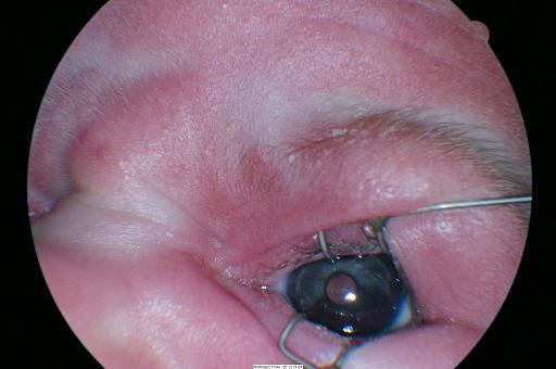
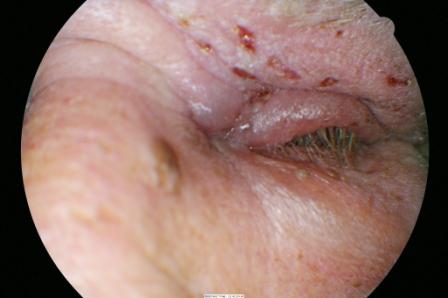
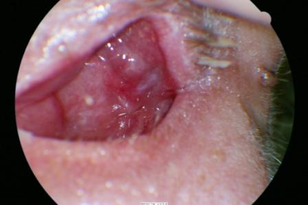
| Periocular petechiae from near asphyxiation |
|
|
|
|
Tuesday, June 16, 2009 |
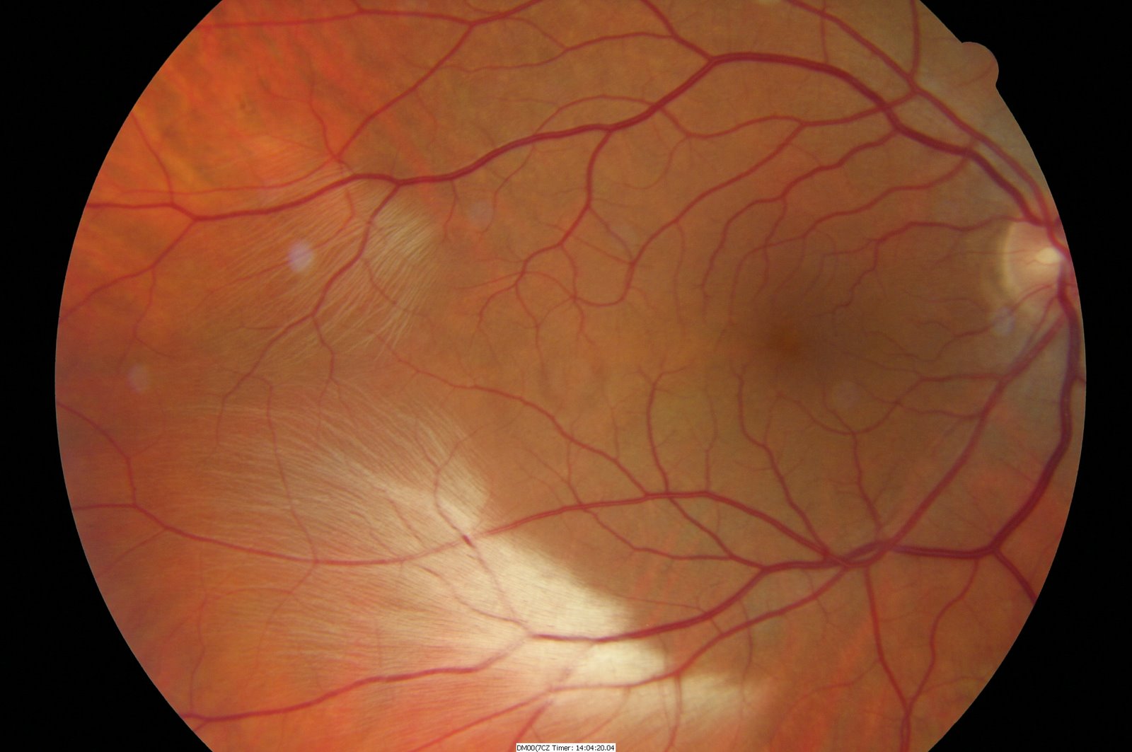
| Retinal break in Stickler's Syndrome |
|
|
|
|
Wednesday, May 6, 2009 |
I'm not sure what fascinates me so much about these dellen from spontaneous subconjunctival hemorrhages. This one is the fourth one I've seen. It cleared up in one day with lubrication. It would be nice to have an anterior segment OCT when these things come by.
|
alsobol at 7:25 AM | | |
Permalink
|
|
|
|
Wednesday, April 29, 2009 |
| A few epiretinal membranes |
|
Not very visually significant but nice images on the OCT.
|
alsobol at 6:59 AM | | |
Permalink
|
|
|
|
Monday, April 27, 2009 |
| Pigment dispersion and farinata |
|
Those farinata are tough to photograph on direct illumination.
|
alsobol at 12:56 PM | | |
Permalink
|
|
|
|
Monday, April 27, 2009 |
Osteogenesis imperfecta. Type I
|
alsobol at 12:53 PM | | |
Permalink
|
|
|
|
Friday, March 27, 2009 |
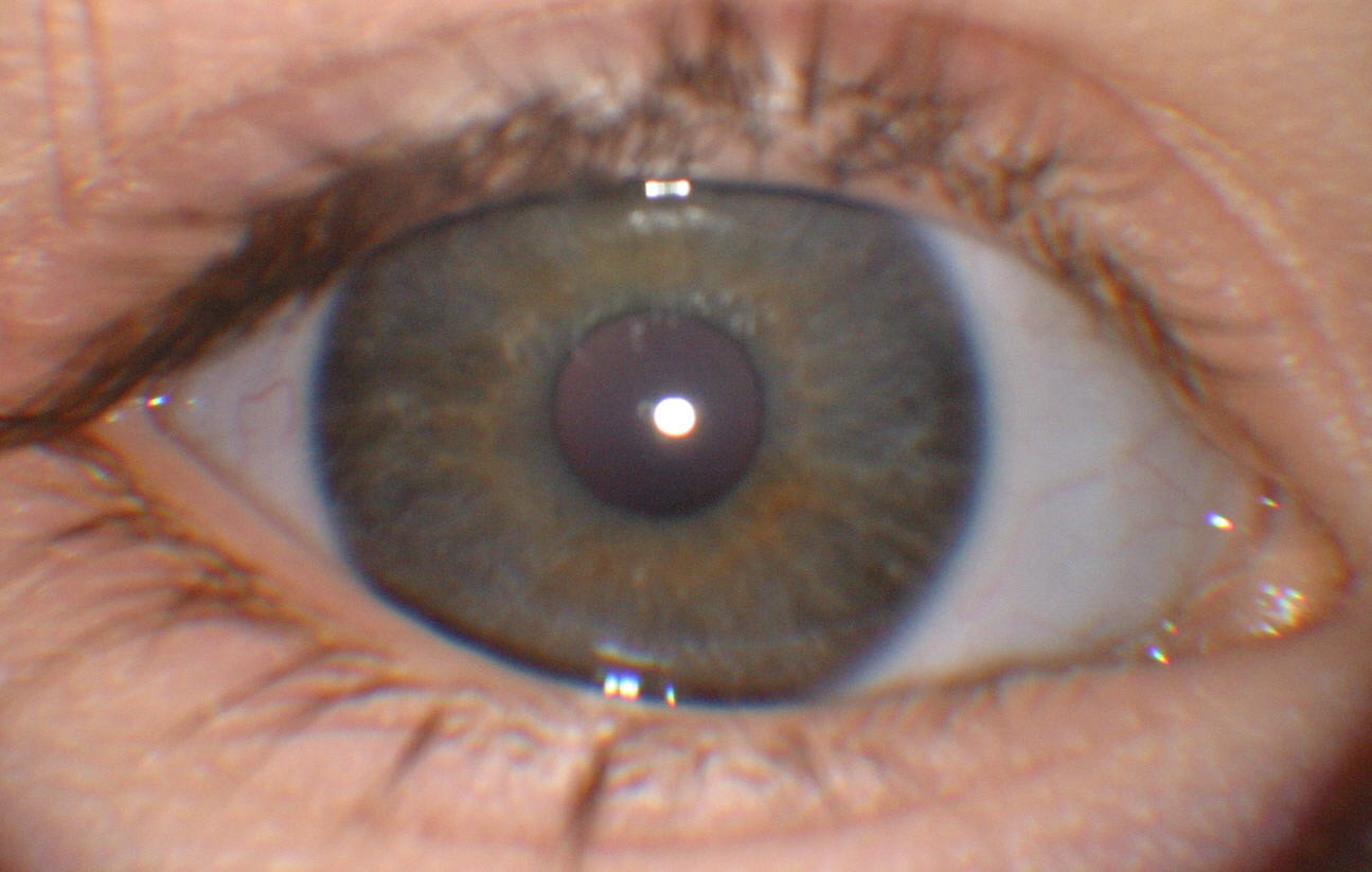
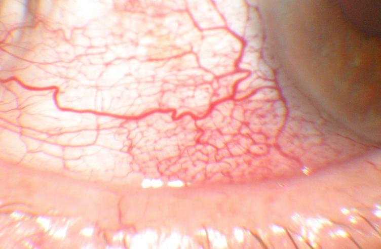
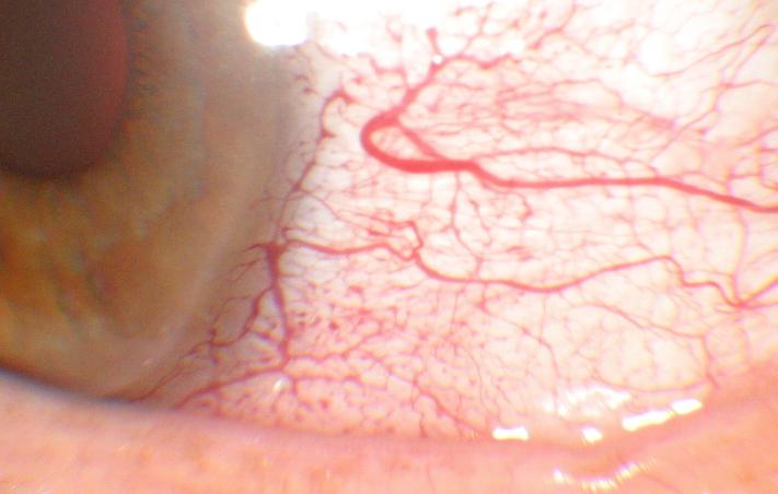
| Conjunctiva in Urticarial Vasculitis |
|
A reticular pattern of conjunctival blood vessels without inflammation.
|
alsobol at 1:40 PM | | |
Permalink
|
|
|
|
Thursday, February 26, 2009 |
Nice photo of pseudoexfoliation of the lens capsule.
|
alsobol at 11:57 AM | | |
Permalink
|
|
|
|
Monday, February 23, 2009 |
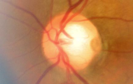
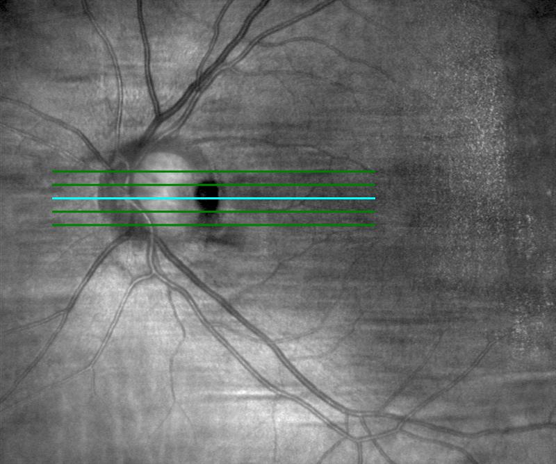
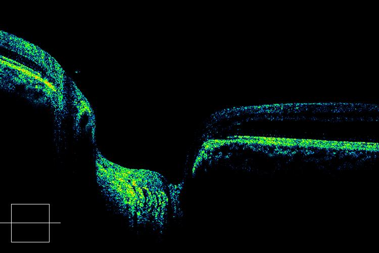
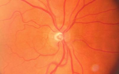
| Optic nerve pit spectral domain OCT |
|
Asymptomatic pit without macular changes. You can see the deep defect in the nerve on the central slice.
|
alsobol at 9:07 AM | | |
Permalink
|
|
|
|
Saturday, February 14, 2009 |
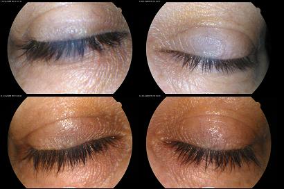
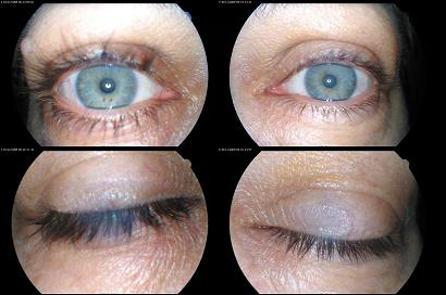
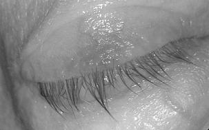
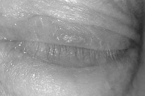
| Update on Lumigan Eyelashes |
|
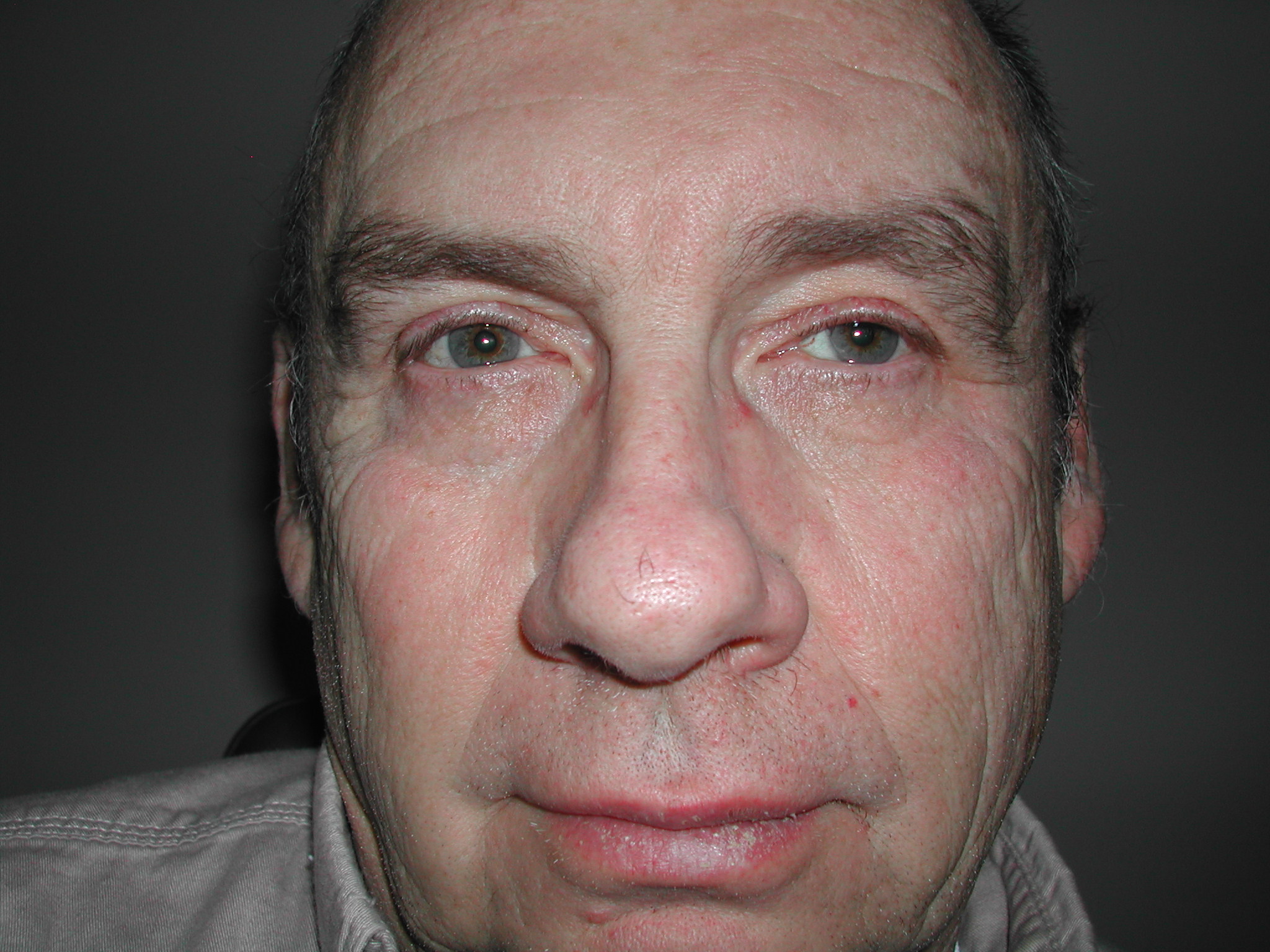
An update on the patient that had asymmetric lashes from monocular Lumigan use and wanted to use it in both eyes for the side effect. Here are her before and after shots: the left eye has much longer lashes now to match the right one.
I think Latisse is going to be pretty popular with results like these.
|
alsobol at 7:08 AM | | |
Permalink
|
|
|
|
Thursday, January 29, 2009 |
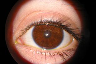
Young woman with 20/20 vision.
|
alsobol at 2:14 PM | | |
Permalink
|
|
|
|
Saturday, January 17, 2009 |
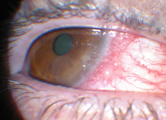
| Nice photo of anterior chamber reaction |
|
Iritis patient
|
alsobol at 9:37 AM | | |
Permalink
|
|
|
|
Saturday, January 17, 2009 |
| Central serous retinopathy on the Cirrus |
|
A new pigment epithelial detachment patient today. I haven't seen one since the Cirrus OCT was installed. It's pretty dramatic. I'm going to try to animate the fluorescein into a .gif file.
|
alsobol at 9:29 AM | | |
Permalink
|
|
|
|
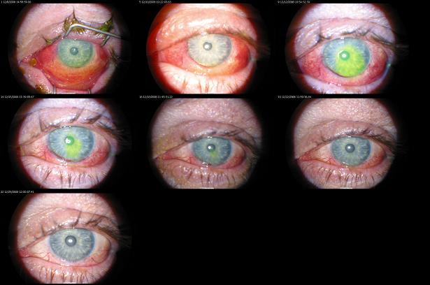
Monday, December 29, 2008 |
Alkali burn with a lot of conjunctival blanching. It's healed slowly but surely over 3 weeks.
|
alsobol at 9:11 AM | | |
Permalink
|
|
|
|
Saturday, December 6, 2008 |
Here's my first try. It works, but I still have to work on the rounding and significant figures. I'll add post-LASIK stuff and some other utilities later. Maybe post a limited version on the appstore without the legal issues.
|
alsobol at 10:26 PM | | |
Permalink
|
|
|
|
Tuesday, November 18, 2008 |
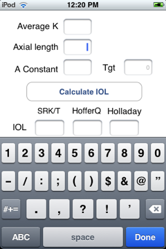
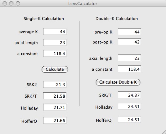
| first step on an iPhone app |
|
I've been working on writing an IOL calculator for the iPhone, but had to learn how to write objective-c first. Here's my first attempt at a program with xcode. I still have to clean up the significant digits and a textbox for target postop refraction. Sadly, all these programs are only for my personal use because of trademark and licensing issues.
|
alsobol at 7:48 PM | | |
Permalink
|
|
|
|
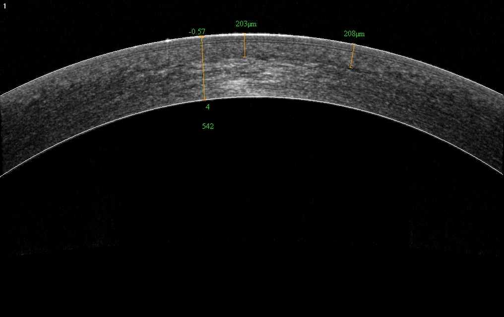
Wednesday, November 12, 2008 |
A nice shot of the LASIK flap on my right eye with the RTVue's flap measuring application.
|
alsobol at 5:49 PM | | |
Permalink
|
|
|
|
Saturday, October 11, 2008 |
I've posted a few times on this but this is the first patient in my practice that wanted to change medications to get her eyelashes to grow. The right eye has been on Lumigan for five or six months and the left eye is well-controlled following SLT. The patient wants to start using Lumigan in her left eye just for the side-effect. Luckily, she isn't having any periorbital skin pigmentation or iris changes.
|
alsobol at 6:55 AM | | |
Permalink
|
|
|
|
Wednesday, October 8, 2008 |
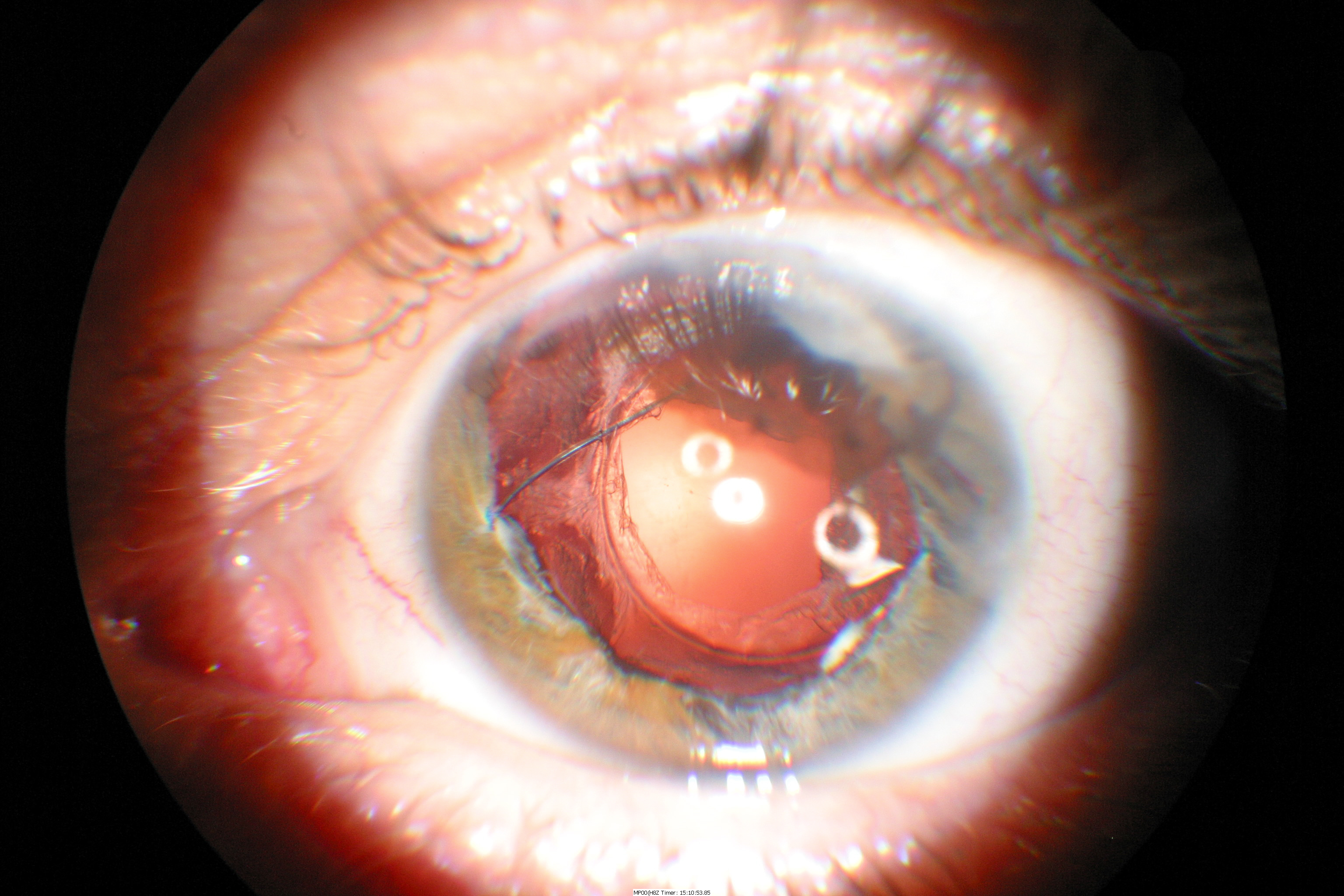
Man, I've had a lot of these recently. The first photo shows a case from last month where I needed SIX hooks to keep the iris in place, and that was with a subincisional hook to control the iris below the wound. You can still see the hexagonal pupil here but it has since resolved to a round pupil. The second photo is straight through the microscope eyepiece with a digital camera, just to see if it could be done; I placed four hooks in a diamond configuation but had to add one under the paracentesis.
|
alsobol at 8:12 AM | | |
Permalink
|
|
|
|
Wednesday, October 8, 2008 |
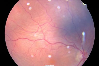
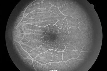
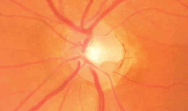
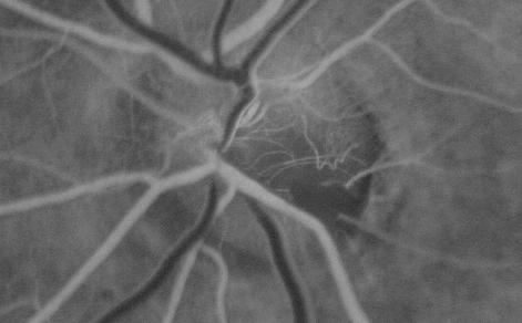
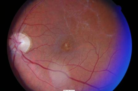
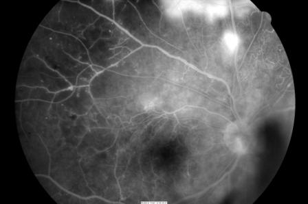
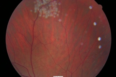
| Combined cilioretinal artery occlusion with central retinal vein occlusion |
|
Rare, but there have been a few case reports and one case series published. I think that the macula has some patchy ischemic retinal whitening (PIRW) in this young patient.
 |
alsobol at 7:56 AM | | |
Permalink
|
|
|
|
Monday, September 22, 2008 |
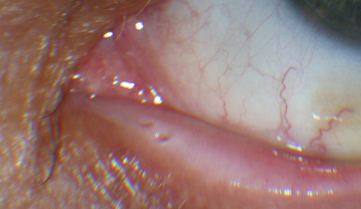
| Subconjunctival hemorrhages causing Dellen formation |
|
Two spontaneous subconjunctival hemorrhages that were elevated enough to cause dellen to form. One of the patients is on coumadin. One patient actually needed a tarsorraphy for exposure. Each was only about 20%.

|
alsobol at 10:36 AM | | |
Permalink
|
|
|
|
Monday, September 22, 2008 |
| Follow-up endothelial changes from chlorpromazine |
|
|
|
|
Monday, September 15, 2008 |
| Pigment Epithelial Detachment |
|
Very dome-shaped PED in this CSR patient. It resolved well spontaneously.
 |
alsobol at 9:18 AM | | |
Permalink
|
|
|
|
Thursday, September 11, 2008 |
| Choroidal striae in two patients |
|
Two patients in just one week.

|
alsobol at 1:42 PM | | |
Permalink
|
|
|
|
Tuesday, September 9, 2008 |
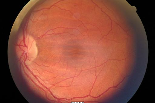
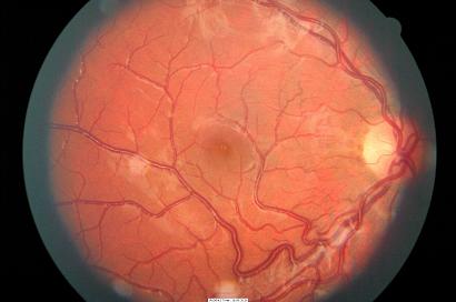
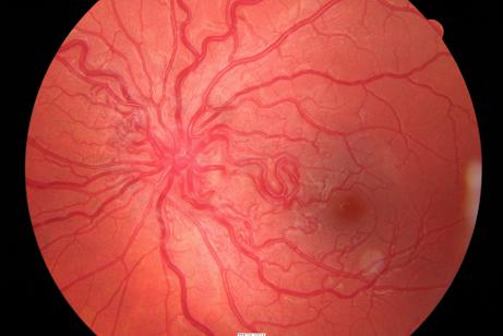
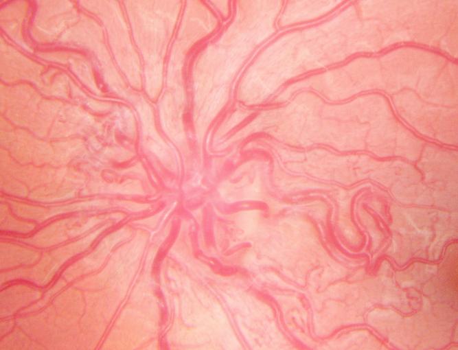
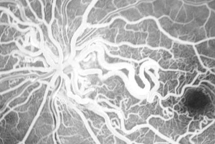
| Congenital retinal macrovessel |
|
I have one other patient where a horizontal macrovessel crosses right through the macula but I think that this vessel almost looks "shocking" because the vessel crosses the horizontal raphe so far vertically.
|
alsobol at 8:40 AM | | |
Permalink
|
|
|
|
Thursday, August 14, 2008 |
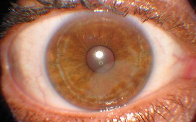
Nice chlorpromazine cataract photo. There is also a fine golden brown pigment dusting on the corneal endothelial surface. It's interesting that the pigment dusting is diffuse and covers the endothelium from white to white and it's not in a vertical spindle like in pigment dispersion syndrome.
|
alsobol at 11:10 AM | | |
Permalink
|
|
|
|
Thursday, August 7, 2008 |
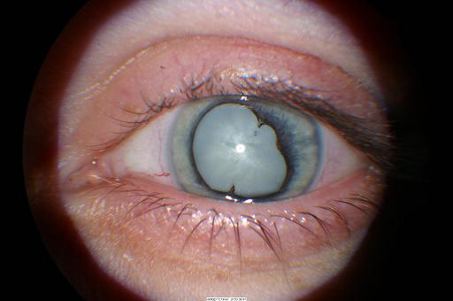
| Phlyctenules and Phacomorphic Glaucoma |
|
Lots of 'ph' words; I saw two phlyctenules today. Here'a a photo of the larger one. The cataractous patient's anterior chamber depth is 2 mm while the fellow eye is 4 mm.
|
alsobol at 12:44 PM | | |
Permalink
|
|
|
|
Tuesday, July 29, 2008 |
| Francois Neetan Dystrophy |
|
Have a couple of patients with this one, but I've never really gotten a good picture of it.
|
alsobol at 2:21 PM | | |
Permalink
|
|
|
|
Friday, July 18, 2008 |
I see this frequently, but on this patient it is only after a short monocular trial of travatan. Here are the eyes: I had to make the photos B/W because the lashes on the left eye are so fine and thin that they can't be seen without the extra contrast.
|
alsobol at 9:32 AM | | |
Permalink
|
|
|
|
Tuesday, July 8, 2008 |
I am not familiar with anything (except trauma) that could cause these interesting linear breaks in Bowman's membrane. It looks very similar to the snail tracks seen in posterior polymorphous membrane dystrophy, except these are very anterior and not in Descemet's membrane. The patient is a young girl with an otherwise unremarkable examination.
|
alsobol at 1:25 PM | | |
Permalink
|
|
|
|
Wednesday, June 25, 2008 |
| Central serous retinopathy |
|
|
|
|
Wednesday, June 25, 2008 |
| NVI and assorted anterior photos |
|
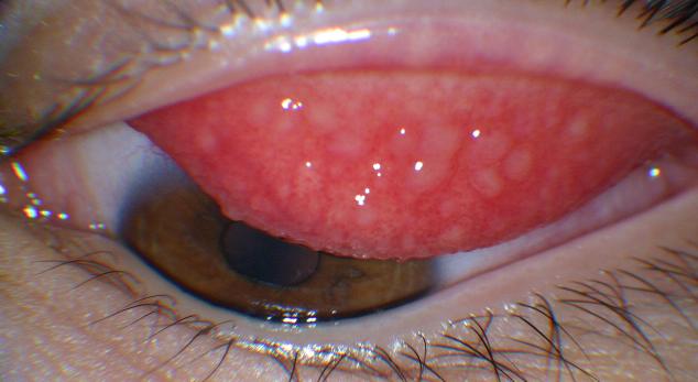
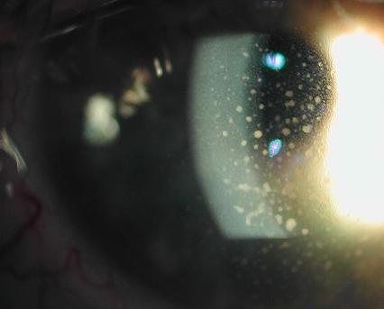
More photos off the slit lamp camera. I must see at least one undiagnosed and asymptomatic EBMD patient daily. GPC is also pretty common locally with our teenagers not changing their disposable lenses. The mutton-fat keratic precipitates are in a sarcoidosis patient.
|
alsobol at 8:08 AM | | |
Permalink
|
|
|
|
Wednesday, June 25, 2008 |
| Double Bowman's, Accessory Punctum, assorted photos |
|
Time to empty the slit lamp camera again. Surpringly there isn't a google image that shows the "double bowman's" after epikeratophakia, even though there are so many of these patients around.
I have four patients with a working accessory punctum. Here are two of them:
|
alsobol at 8:00 AM | | |
Permalink
|
|
|
|
Friday, June 6, 2008 |
Blunt trauma with iritis, angle recession, a few retinal tears, and a central choroidal rupture. The second photo shows it a few days afterwards where there is a small choroidal hemorrhage and the third photo shows the fluorescein that shows no (early) choroidal neovascularization causing the hemorrhage.
|
alsobol at 8:58 AM | | |
Permalink
|
|
|
|
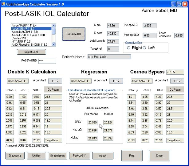
Wednesday, June 4, 2008 |
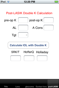
| Great IOL selection in post-LASIK patient |
|
Always a nail-biter for IOL power selection in these post-LVC patients. I just saw a patient for her POD #1 visit and she was 20/20, a huge relief. I had all of her pre-and-post procedure records available and was able to run the DoubleK, Corneal bypass, Feiz-Mannis, and Masket calculations for S*K/T, Hoff**Q, and Holl**ay1. Here's a screenshot from a Visual Studio program that I wrote a few years ago to run the calculations.
|
alsobol at 5:47 PM | | |
Permalink
|
|
|
|
Friday, April 25, 2008 |
Two recent insect-related eye injuries. The first one is a recent patient with a hyphema after the bee smacked him in the eye.
The next two photos were from a patient who was stung in the cornea by a bee; the patient notes that he is allergic to bee stings. The stinger is visible paracentrally and there was dramatic corneal whitening and edema. The second photo shows the healed cornea following removal. Note: I get no credit for the excellent result, WVU Corneal Service removed the stinger.
|
alsobol at 1:18 PM | | |
Permalink
|
|
|
|
Friday, April 25, 2008 |
| Resolution of Sectoral Heterochromia, sort of |
|
Neonate that I saw for an iris lesion consistent with sectoral heterochromia. On a follow-up exam (more than 3 years later) the remainder of her iris has darkened and the heterochromia is no longer evident.
|
alsobol at 10:02 AM | | |
Permalink
|
|
|
|
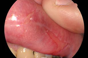
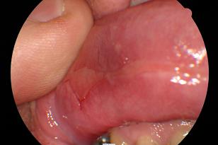
Thursday, April 17, 2008 |
Nice guy with painful oral ulcers for two months and conjunctivitis for three weeks. 26 pound weight loss from the dysphagia. buccal sample sent for direct immunofluorescence - was positive. (that's the patients finger in the photo, not mine)
update 4/17/2008
buccal and conj sample positive for strong IgG epithelial cell deposition
|
alsobol at 9:06 AM | | |
Permalink
|
|
|
|
Wednesday, April 2, 2008 |
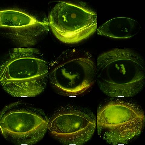
| HSV epithelial keratitis collage |
|
Some more photos from the transfer:
|
ALSOBOL at 7:03 PM | | |
Permalink
|
|
|
|
Wednesday, April 2, 2008 |
Transferred my old photos to my new laptop. Here's a few.
|
ALSOBOL at 6:55 PM | | |
Permalink
|
|
|
|
Thursday, March 27, 2008 |
Fine discrete corneal crystals from limbus to limbus.
Friday, April 04, 2008
update: M spike on SPEP, gamma on immunofixation.
|
alsobol at 1:13 PM | | |
Permalink
|
|
|
|
Tuesday, March 25, 2008 |
Unusual to see two of these in just two weeks.
|
alsobol at 2:02 PM | | |
Permalink
|
|
|
|
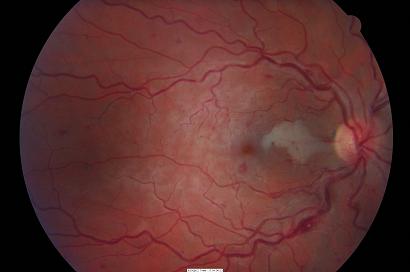
Thursday, March 20, 2008 |
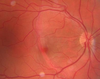
| Retinal finding following hyphema |
|
Young woman with an unusual deep retinal lesion (hemorrhage?) following a hyphema. She has 180 degrees of angle recession and some iris sphincter damage but otherwise no problems.
Can you see it? It's easier to see on red-free and fluorescein:
|
alsobol at 10:49 AM | | |
Permalink
|
|
|
|
Tuesday, March 18, 2008 |
| Bilateral interstitial keratitis |
|
Young woman with bilateral deep scarring with ghost vessels. Not usually an interesting case, but she presented today for a second opinion on a corneal transplant. She was told by the another corneal specialist that she should have bilateral simultaneous corneal transplants. She is 20/25+ in both eyes.

|
alsobol at 11:55 AM | | |
Permalink
|
|
|
|
Saturday, March 15, 2008 |
Centrally located optic nerve pit without macular changes.
|
alsobol at 9:14 AM | | |
Permalink
|
|
|
|
Thursday, March 6, 2008 |
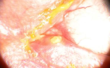
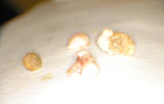
| Gemstones of the lacrimal duct |
|
Dacryoliths expressed from this patient with intermittent symptoms.
|
alsobol at 1:43 PM | | |
Permalink
|
|
|
|
Thursday, March 6, 2008 |
Ectodermal dysplasia with limbal stem cell deficiency, corneal scarring, and sensory nystagmus. Similar presentation in his brother; mother and aunt with less severe form.
|
alsobol at 1:42 PM | | |
Permalink
|
|
|
|
Wednesday, February 27, 2008 |
| Resolution of bitemporal hemianopsia over two years |
|
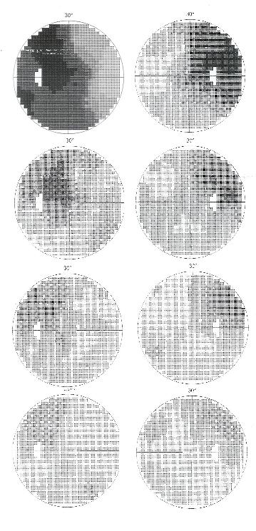
Two years ago I found a pituitary macroadenoma in this very pleasant 83 year-old lady (based on confrontational visual fields and left optic neuropathy). She had it resected and slowly recovered her full fields over two years. It is impressive that she is still continuing to improve; the first visual field is her baseline two years ago, the second at four months, the third at eight months, and the last at twenty months.
|
alsobol at 5:20 PM | | |
Permalink
|
|
|
|
Tuesday, February 12, 2008 |
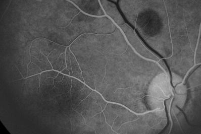
Racemose hemangioma found on routine exam with otherwise normal 20/20 vision. The dilated retinal vein has no capillaries around it, as seen on this
angiogram. Also, You can see that it fills very quickly, much sooner
than the other retinal veins that are just starting to fill.
|
alsobol at 1:24 PM | | |
Permalink
|
|
|
|
Wednesday, February 6, 2008 |
Miller Fisher variant of Guillain Barre Syndrome. Patient presented with ataxia and was admitted for workup. Here's a video of his ophthalmoplegia from my cell phone when I first met him. He got IVIG for a few days and was discharged.
I lost him for a month but he came back today with his EOM's dramatically improved and walking well. He also was admitted to a local tertiary care center recently but I'm not sure if they gave him any more IVIG or plasmapheresis.
This videos won't run in Quicktime and have to run in Windows Media Player.
|
alsobol at 8:00 PM | | |
Permalink
|
|
|
|
Tuesday, February 5, 2008 |
| Iris in neovascular glaucoma |
|
I'm getting pretty good with these iris angiograms. This patient presents with neovascular glaucoma from proliferative diabetic retionpathy.
|
alsobol at 1:40 PM | | |
Permalink
|
|
|
|
Tuesday, January 22, 2008 |
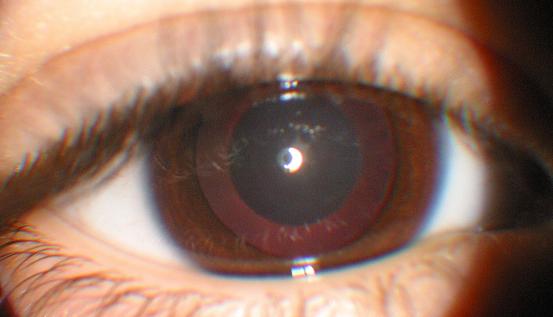
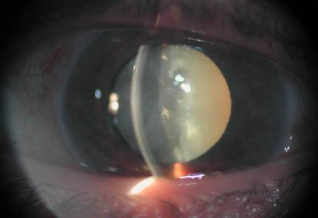
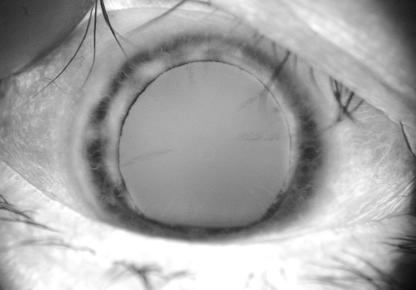
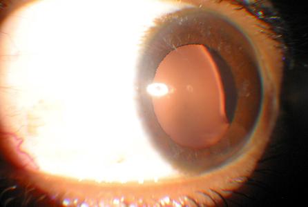
Nice guy punched in the eye thirty years ago. He has no zonules over the entire temporal edge of the lens with phacodonesis and a large bubble of vitreous in the anterior chamber.
Don't see too many of these and it is interesting to compare the lens edge with the patient from a few posts ago. This mature lens dislocates and the lens edge is smooth. The lens edge is flat if an immature lens dislocates.
 |
alsobol at 12:38 PM | | |
Permalink
|
|
|
|
Thursday, January 17, 2008 |
fluorescein angiogram of iris neovascularization from proliferative diabetic retinopathy. I think I'm going to start to take an iris photo routinely when I know the patient has new retinal vessels.
|
alsobol at 10:56 AM | | |
Permalink
|
|
|
|
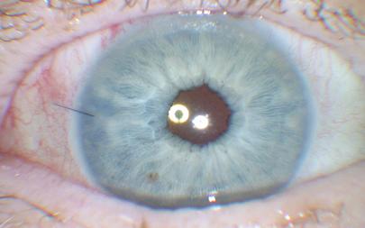
Friday, January 11, 2008 |
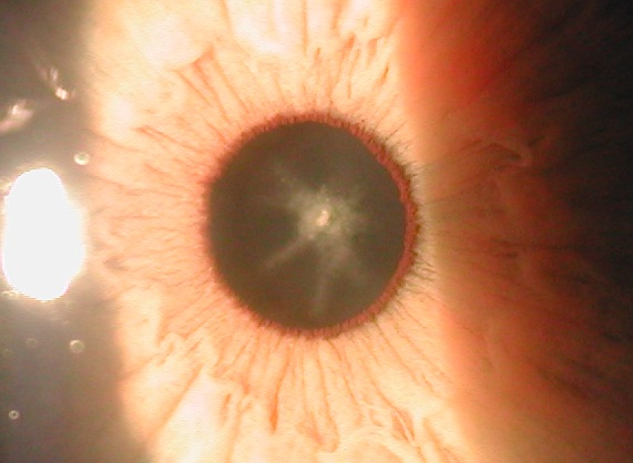
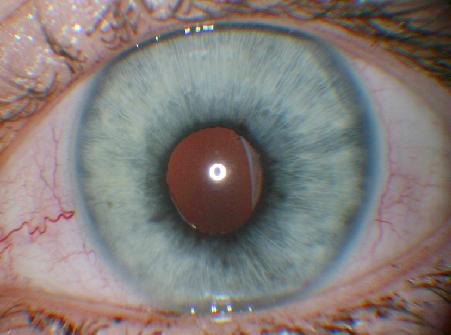
| nice pigment dispersion syndrome retroillumination photo |
|
|
|
|
Friday, January 11, 2008 |
very tall six year-old with lenticular astigmatism. serum and urine studies pending.
|
alsobol at 1:25 PM | | |
Permalink
|
|
|
|
Wednesday, January 9, 2008 |
| small incision levator advancement (in office) |
|
Dr. Hormozy taught me how to do this in my residency (drhormozy.com). In the office, you make a 3mm incision at the upper tarsal border, dissect superiorly until you can grasp the levator with forceps, and then suture it to the lower tarsus. The problem with the small single incision is that you end up with a "peak" centrally in the eyelid. This patient's other eyelid has a natural "peak" so she is a good candidate; also, she doesn't want to go to the OR. You can make a larger incision to have a better contour but then you may as well just do it in the OR so you can remove the fat pads, etc..
You can be sure that you have a good grip on the levator (you can't see it) when the suture is dragged superiorly when the patient is asked to look up.
|
alsobol at 5:17 PM | | |
Permalink
|
|
|
|
Thursday, December 20, 2007 |
A patient had a penetrating injury thirty years ago with loss of his superior iris and diffuse synechiae OS. He had a dense dislocated cataract. I took out the cataract and attached the haptics to the stubs of his iris at 3 and 9 o'clock with 10-0 prolene. This is an old case prior to capsular rings. He is pretty happy with the result. He has some cylinder from the large superior corneal scar and he had a YAG capsulotomy last year. I regret not taking a pre-op photo. Here he is today at 20/30:
|
alsobol at 12:23 PM | | |
Permalink
|
|
|
|
Tuesday, December 18, 2007 |
| Longstanding aberrant regeneration |
|
On routine exam found a man who had presumed left blepharospasm for 37 years. It can be isolated to when he moves his jaw to the left. He will return for botox soon although he doesn't think it will work as he has tried many modalities over the past decades.
Here he is with his jaw moved laterally to the left. He has unrelated XT and amblyopia OS.
|
alsobol at 1:29 PM | | |
Permalink
|
|
|
|
Wednesday, November 21, 2007 |
| Slit lamp camera: Avellino dystrophy and Haab's Striae |
|
I bought a camera adapter for my Nikon 880 that fits into the slip-lamp eyepiece. The pictures are dark unless you also attach a separate lamp, but I think they still show the photos pretty well.
Here are a few good ones.
Avellino corneal dystrophy
Haab's striae
|
alsobol at 7:38 AM | | |
Permalink
|
|
|
|
Wednesday, November 21, 2007 |
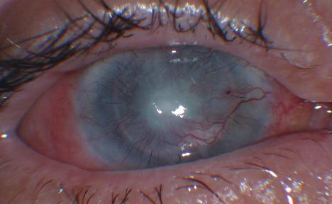
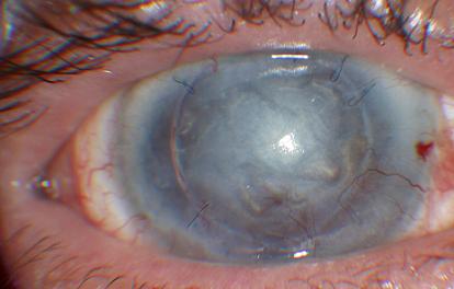
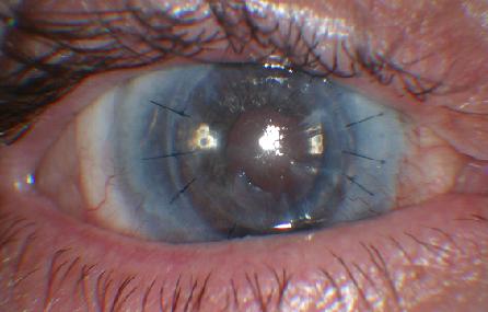
A patient with a perforating injury forty years ago and an updrawn and phimotic pupil with posterior synechiae. He was nine years old when it occurred and he was game to try and repair it. We planned to break the synechiae with Kuglen hooks and Healon in the OR and then enlarge and move the pupil later via laser iridoplasty. Here he is pre-operatively:
And this morning. There is some corneal edema at one of the paracentesis sites; he likely has some endothelial loss from the injury. Luckily there is no cataract.
Wednesday, November 28, 2007
UPDATE
Things are healing well and the corneal edema has essentially resolved. He's 20/400 uncorrected and I haven't tried to refract him yet. His lens is clear and only looks hazy in the photo; he probably has a lot of cylinder from the scar. Here's the one week follow-up:
|
alsobol at 7:28 AM | | |
Permalink
|
|
|
|
Saturday, November 17, 2007 |
| Zoster following enucleation |
|
I never thought about it before but there really isn't any reason that enucleation would stop zoster conjunctivitis in HZO (orbit-itis?)
|
alsobol at 7:25 PM | | |
Permalink
|
|
|
|
Saturday, November 17, 2007 |
Hypertensive retinopathy with old BRVO with non-perfused scarred macula. The interesting part, the retinal vein crosses over the artery. I probably wouldn't have noticed it unless there was other pathology.
Here's a close-up of the crossing:
Update: Friday, January 25, 2008
Actually, I've seen this again since I started looking for it. It must be more common than I thought.
|
alsobol at 7:06 PM | | |
Permalink
|
|
|
|
Monday, October 22, 2007 |
Early last year I saw a patient with a total carotid occlusion for the past 10 years. She had an ischemic retinopathy with tortuous retinal vessels. Here she was 18 months ago:
She presented today with an ischemic central retinal vein occlusion.
The other carotid was clean at last check and a repeat doppler is pending.
|
alsobol at 4:53 PM | | |
Permalink
|
|
|
|
Sunday, October 21, 2007 |
A case of bartonella neuroretinitis with a nice macular star and papillitis.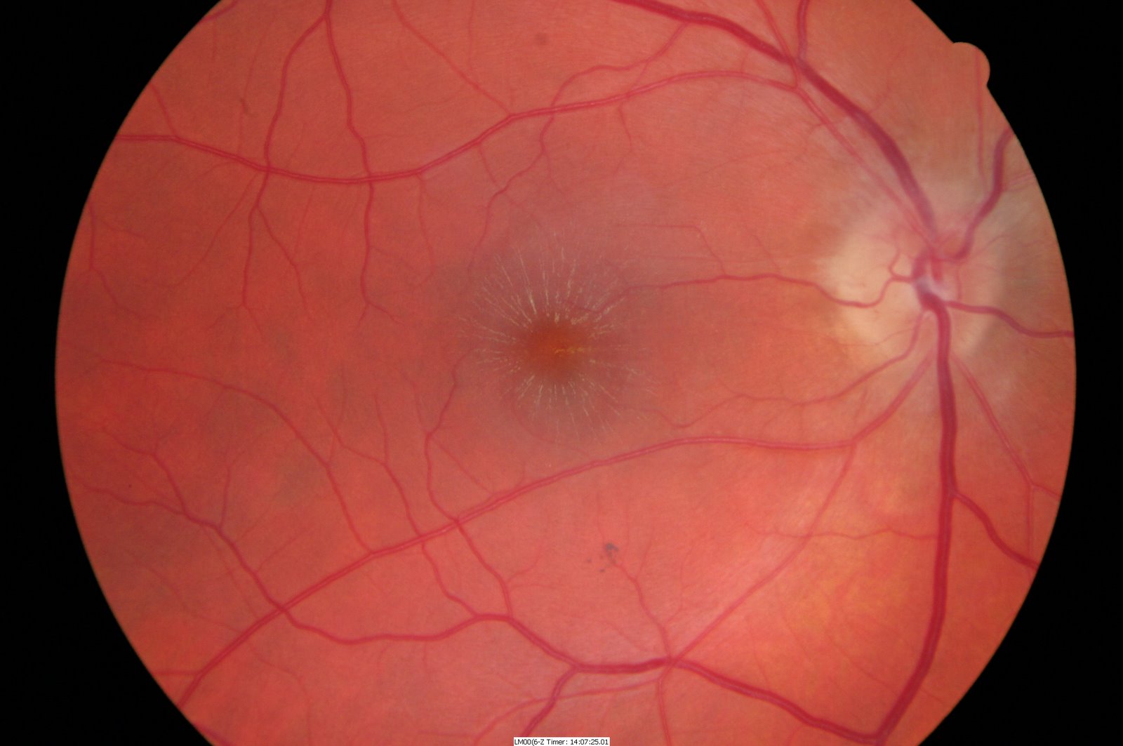
It cleared well over a few weeks with oral antibiotics:
The vet said that for cat scratch disease the cat only needs good flea control, no need for antibiotics for the cat.
|
alsobol at 6:55 PM | | |
Permalink
|
|
|
|
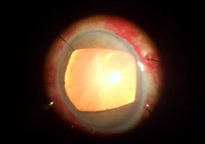
Thursday, October 4, 2007 |
| Anterior capsule in liquified uveitic cataract |
|
I had a follow-up today of a patient with idiopathic bilateral panuveitis who had a white liquified cataract. Here she is pre-operatively.
I was unable to get the anterior capsule to bend over itself to begin the capsulorrhexis, so I just aspirated the cataract through a small linear incision in the anterior capsule. I extended the tear to 12 and 6 o'clock and oriented the IOL so that the haptics were oriented horizontally and "pushed" the anterior leaflets of the anterior capsule toward the limbus.
Here she is on POD #1. Note how the anterior capsular leaflets are almost appositional. Over the next week she had a flare-up including a dense vitritis and papillitis. This all resolved with oral steroids and a sub-Tenon's injection. You can see the inflammed vessels on her iris.

Here she is today; the leaflets spread away nicely, probably due to tension from the IOL haptics.
10:33:48 AM Sunday, October 07, 2007
Addendum:
I found a better way to do it:
ceiol.com or
http://www.yousendit.com/transfer.php?action=batch_download&batch_id=cUJVc2ZBYTIzMW5IRGc9PQ
|
alsobol at 11:33 AM | | |
Permalink
|
|
|
|
Tuesday, October 2, 2007 |
| One of my favorite pictures |
|
Myelinated nerve fibers showing the orientation of the nerve fiber layer.
|
alsobol at 5:01 PM | | |
Permalink
|
|
|
|
Tuesday, October 2, 2007 |
| Guess which eye is on Lumigan? |
|
|
|
|
Tuesday, October 2, 2007 |
A patient with severe eczema and chronic corneal irritation had two prior transplants OD and one OS. Presented two years ago with severe vascularization with opacification OU and he had poor healing and chronic epithelial defects after the first grafts. The left eye also had a perforation. Hand motions/light perceptions pre-op.
Here's his pre-op photo. I gave him a limbal stem cell allograft. Dr. Tseng's lab in Miami did the limbal impression cytology.
Here he is 6 months after the allograft, the grafts are at 3 and 9 o'clock. Note the smooth and avascular corneal epithelium. I placed the grafts in the interpalpabral fissure for cosmesis.He was lost to follow-up immediately after the surgery and did not get any immunosuppressants. The graft survived well.
Then the new corneal graft.
The photo is out-of focus but you can see the cornea is clear centrally. The patient was lost to follow-up (again) for six months after the PKP and there is some haze in the graft from 3 to 6 o'clock but he is happy with his vision. The epithelium has survived two years without immunosuppressives. Best vision is (I think) 20/80.
He has a similar presentation OS and I gave him a stem cell transplant in that eye five months ago. Here's the post-op photo OS. We'll go for a triple procedure soon. This one will be more challenging as there is a white liquid cataract and a very shallow chamber with anterior synechiae to the peripheral cornea from his prior perforation.
That's keratinized conjunctiva temporally.
|
alsobol at 12:54 PM | | |
Permalink
|
|
|
|
Tuesday, October 2, 2007 |
| A case from this morning. |
|
|
|
|
Thursday, September 13, 2007 |
A small contact-lens associated corneal ulcer, but there was a lot of inflammation including fibrin in the a/c and some posterior synechiae. Turned-out pretty well. 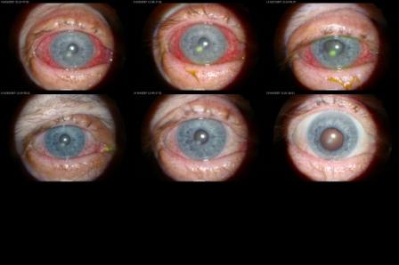
|
alsobol at 7:49 PM | | |
Permalink
|
|
|
|
Monday, September 10, 2007 |
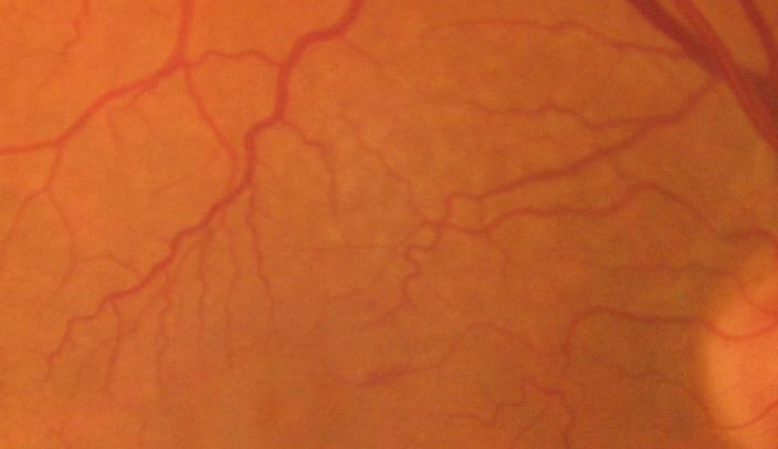
No complaints or other findings in this patient. Congenital tortuous vessels?
|
alsobol at 6:54 PM | | |
Permalink
|
|
|
|
Friday, September 7, 2007 |
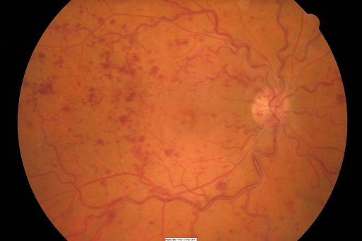
An unfortunate patient who presented for her first diabetic eye exam. The angiogram tells the story pretty well.
|
alsobol at 10:27 AM | | |
Permalink
|
|
|
|
Friday, September 7, 2007 |
Here's a retinopexy from a few weeks ago for a small horseshoe tear. It's not really interesting but it's a good photo for such a peripheral lesion.
|
alsobol at 10:04 AM | | |
Permalink
|
|
|
|
Wednesday, September 5, 2007 |
I updated the site to Jeremy Wadsworth's template version 2.8 and lost the last month of blog entries, so I'll upload a few of the cases that were up on the old site. I'm having some problems with the masterpages but all the programs are still working.
|
alsobol at 8:25 PM | | |
Permalink
|
|
|
|